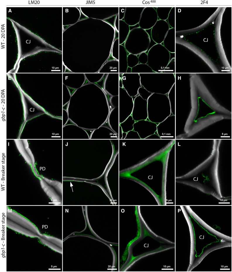
Figure 6.
Cell Wall Alterations in the gbp1-c Mutant.
(A) to (P) Indirect immunofluorescence microscopy of the wild type (WT) and gbp1-c at 20 DPA or at the breaker stage.
Indirect immunofluorescence microscopy was performed using antibodies and probes that indicate different degrees of esterification of the HG component of pectin, from highly esterified (LM20 antibody) to partially esterified (JIM5 antibody, COS488), including no-esterified forms that are able to bind to cations such as calcium (Ca2+) and form gels in the middle lamella (2F4 antibody). Green represents specific antibody or probe signal. Arrow indicates JIM5 signal close to the plasma membrane. The white signal shows Calcofluor white staining of cell walls (β-linked glucans: callose and cellulose). CJ, cell junctions; PD, plasmodesmata.

