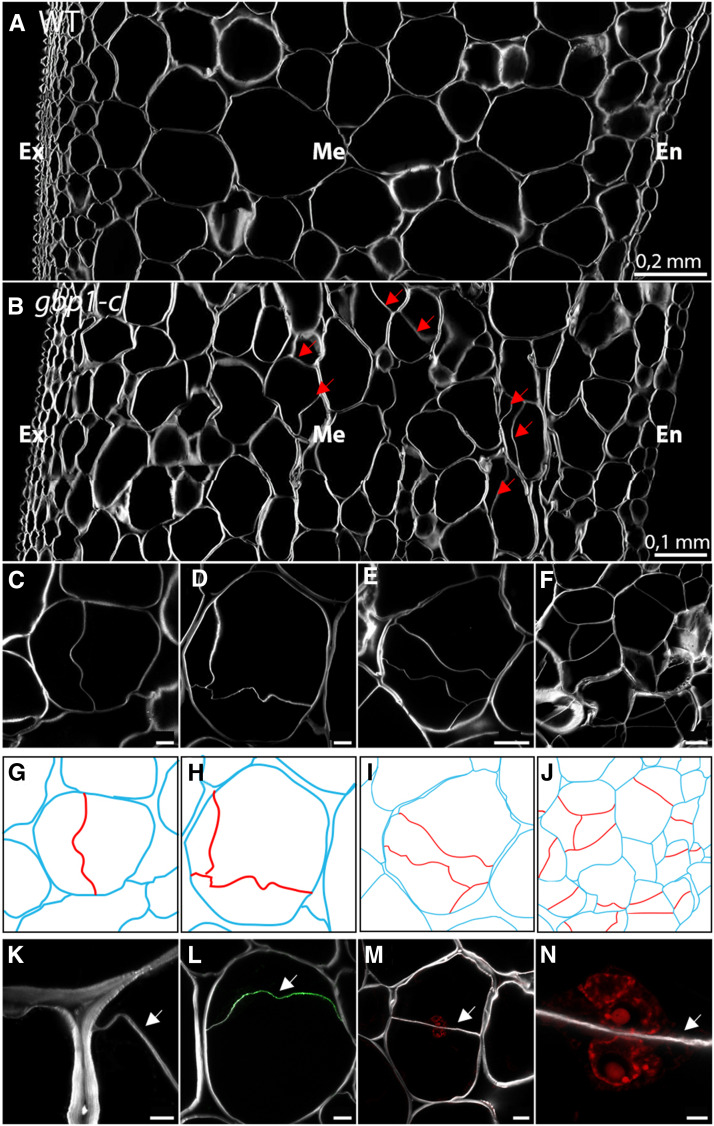
Figure 7.
Newly Formed Cell Walls in Large Mesocarp Cells of the gbp1-c Mutant at the Breaker Stage.
(A) and (B) Pericarp sections in the wild type (WT) and gbp1-c at the breaker stage.
(C) to (F) Additional cell walls inside mesocarp cells in the gbp1-c mutant.
(G) to (J) Schematic representations of cells shown in (A) to (D). The parental cell walls are shown in blue and additional cell walls are shown in red.
(K) Close-up of fusion site between parental and new cell wall.
(L) Indirect immunofluorescence with an anti-β-1,3-glucan antibody (green signal) indicating callose deposition.
(M) and (N) Newly divided parental mesocarp cell showing a nucleus in both daughter cells. Nuclei in red are labeled with propidium iodide.
Scale bars represent 5 µm in (K) and (N), 20 µm in (C), (D), (L), and (M) and 50 µm in (E) and (F).
The white signal shows Calcofluor white staining of cell walls (β-linked glucans: callose and cellulose). Arrows indicate new cell walls. Ex, exocarp; M, mesocarp; En, endocarp.

