Figure 2.
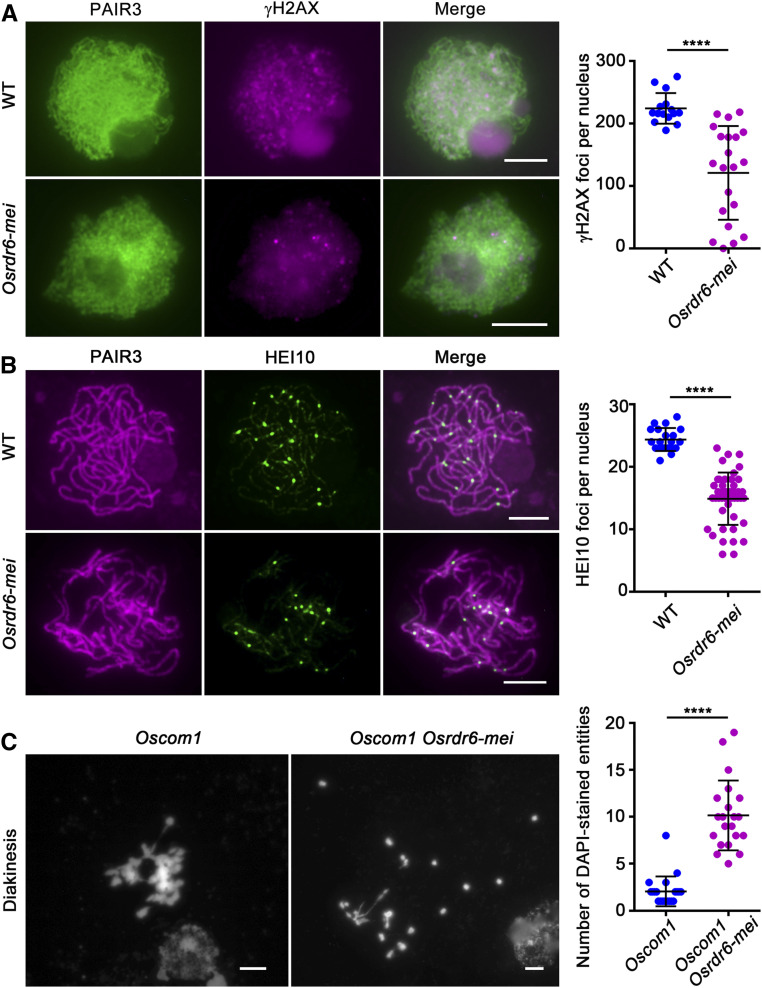
Cytological Analysis of Osrdr6-mei.
(A) Immunofluorescence imaging of γH2AX signals on meiotic chromosome spreads of wild-type (WT) and Osrdr6-mei meiocytes. ****P < 0.0001, two-tailed Student’s t test.
(B) Dual immunolocalization of PAIR3 (red) and HEI10 (green) in wild-type (WT) and Osrdr6-mei meiocytes. PAIR3 was used to visualize the meiotic chromosomes. ****P < 0.0001, two-tailed Student’s t test.
(C) Meiotic chromosome behavior in Oscom1 and Oscom1Osrdr6-mei. Chromosomes were stained with DAPI. Numbers of DAPI-stained entities per cell at diakinesis in Oscom1 and Oscom1Osrdr6-mei plants were analyzed. ****P < 0.0001, two-tailed Student’s t test. Bars = 5 μm.

