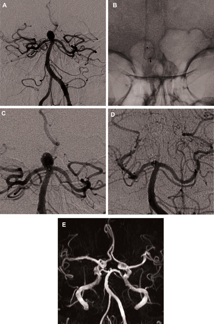
FIGURE 2.
Unruptured basilar tip aneurysm (mean transverse diameter: 6.3 mm; mean height: 6.7 mm; neck: 4.3 mm). A, DSA (frontal view) shows the aneurysm. B and C, Postoperative DSA (unsubstracted and subtracted view, respectively) show the detached WEB device (WEB SL 7 × 4 mm) and residual flow in the aneurysm and the device. D, One-year DSA shows complete aneurysm occlusion. E, Two-year MRA shows stable complete aneurysm occlusion.

