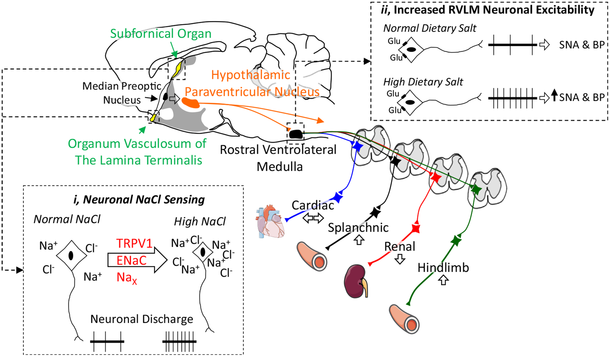
Figure 1.

Dietary salt alters autonomic function by excitation of NaCl-sensing neurons in the lamina terminalis or increasing gain/excitability of bulbospinal neurons in the rostral ventrolateral medulla. Salt-sensitive hypertension is associated with increased sympathetic outflow to the splanchnic or hindlimb vasculature. A midsagittal section of the rodent brain illustrates key autonomic centres involved in salt-sensitive hypertension. (i) Neurons in the organum vasculosum of the lamina terminalis and subfornical organ sense changes in extracellular NaCl to increase sympathetic nerve activity. Potential NaCl-sensing mechanisms include an N-terminal variant of the transient receptor potential cation channel subfamily V member 1 (TRPV1), the epithelial sodium channel (ENaC), and the Nax channel. (ii) Dietary salt also increases the excitability or gain of bulbospinal sympathetic neurons of the rostral ventrolateral medulla. Thus, glutamatergic (or GABAergic) input onto RVLM neurons results in an exaggerated discharge and change in sympathetic nerve activity. BP, blood pressure; RVLM, rostral ventrolateral medulla; SNA, sympathetic nerve activity. Adapted from Servier Medical Art by Servier licensed under a Creative Commons Attribution 3.0 Unported License (https://smart.servier.com).
