Figure 10.

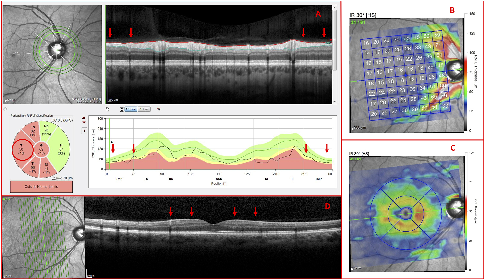
Vertical b-scans verify glaucomatous thinning (A) Inner circle scan, (B) RNFL thickness map, (C) GCL thickness map, and (D) vertical meridian b-scan of a G Eye carrying glaucomatous temporal (T) RNFL thinning (red arrows) and GCL thinning

Vertical b-scans verify glaucomatous thinning (A) Inner circle scan, (B) RNFL thickness map, (C) GCL thickness map, and (D) vertical meridian b-scan of a G Eye carrying glaucomatous temporal (T) RNFL thinning (red arrows) and GCL thinning