
Figure 4.
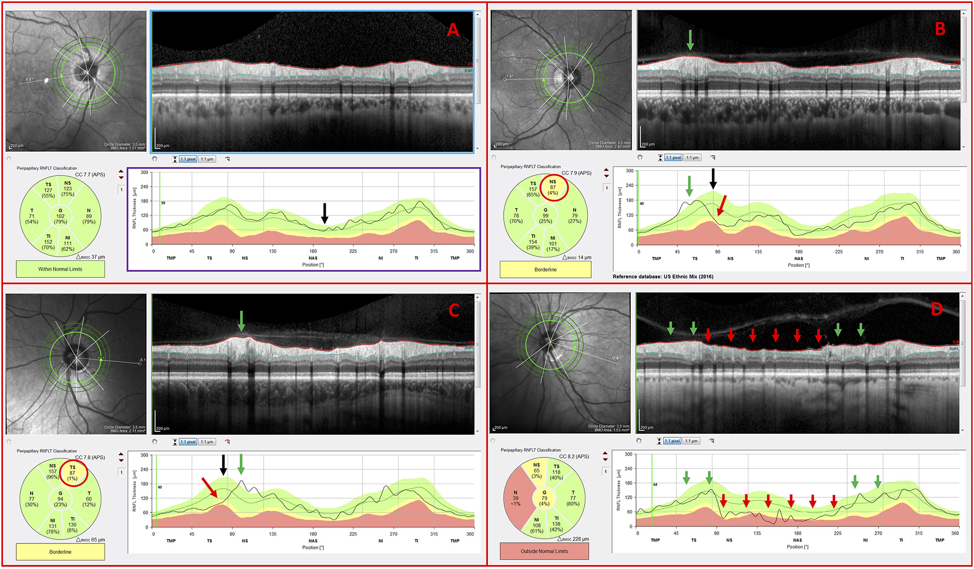
Inner circle scans of non-glaucomatous (NG) eyes carrying the following observed features: (A) cpRNFL thickness profile within normal limits (B) blood vessel displacement toward the temporal region, (C) blood vessel displacement toward the nasal region, and (D) NG optic neuropathy