
Figure 6.
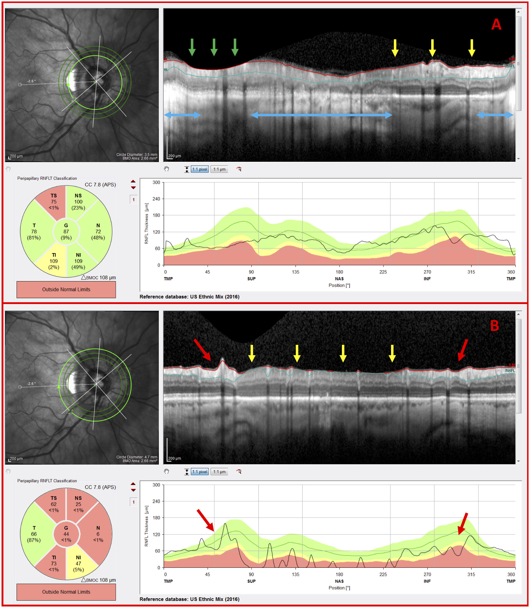
G eye with disruptive co-existing pathology and OCT artifacts (A) Inner circle scan and (B) outer circle scan of a G eye carrying peripapillary atrophy (PPA) (blue arrows), a clipping artifact (green arrows), regions of inadequate contrast (yellow arrows), and indications of glaucomatous damage (red arrows)