-
A, B
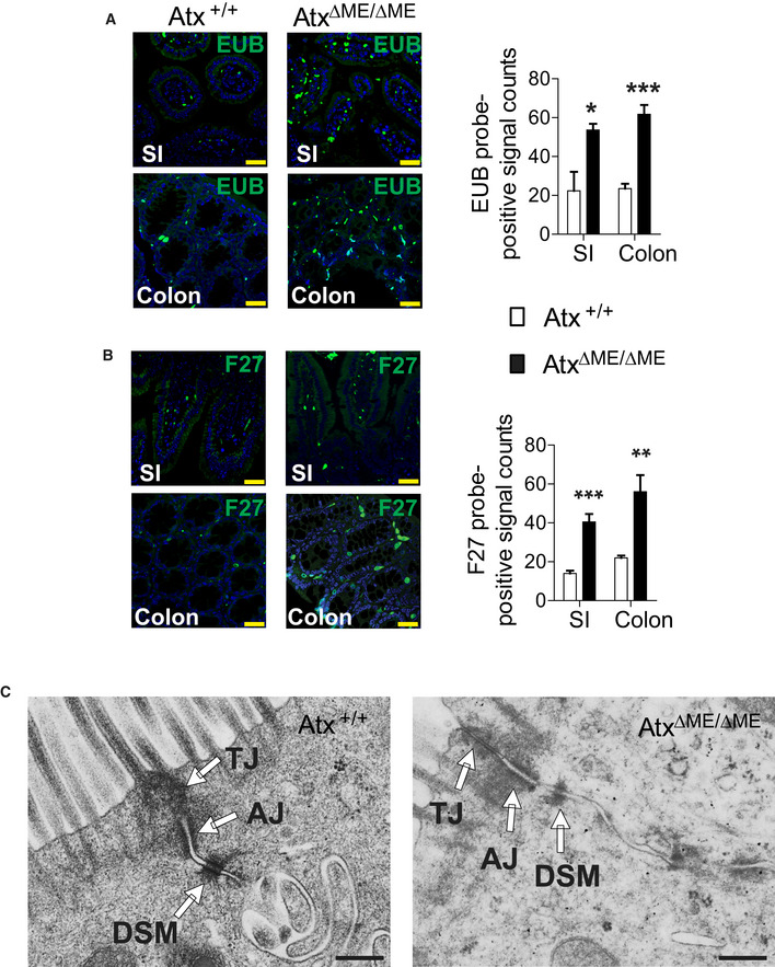
The bacterial load in the intestinal mucosa of AtxΔΜΕ/ΔΜΕ and Atx+/+ mice was evaluated by Fluorescent In Situ Hybridization (FISH) using FITC‐labeled pan‐bacterial EUB probe (A) and FITC‐labeled pan‐bacterial F27 probe (B). Scale bar is 30 μm. Graph indicates the count of FITC‐positive signals quantified from three or more independent experiments (n = 3–4 per group). The data are shown as mean ± SEM. Small Intestine (SI).
-
C
Electron micrographs of intact cell‐to-cell adhesion in the colonic epithelium from age (8 weeks)‐matched AtxΔΜΕ/ΔΜΕ mice and Atx+/+ littermate. Tight junction (TJ), Adherens junction (AJ), Desmosome (DSM). Scale bar indicates 500 nm.
Data information: All images presented are the representative from at least three independent experiments. *
P < 0.05, **
P < 0.01, ***
P < 0.001 (one‐tailed unpaired
t‐test).

