-
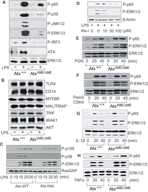
A–C
The macrophages from AtxΔΜΕ/ΔΜΕ and Atx+/+ mice (A, B) or Atx‐heterozygous (Atx‐Het) mice and wild‐type littermates (C) were stimulated by LPS [20 ng/ml, 20 min (A, B) or indicated time point (C)].
-
D
Raw264.7 cells were stimulated by LPS with the ATX inhibitor.
-
E–H
The macrophages from AtxΔΜΕ/ΔΜΕ mice and Atx+/+ littermates were activated with TLR2 ligand Peptidoglycan (PGN, 30 ng/ml) (E), Pam3CSK4 (1 μg/ml) (F), IL‐1β (100 ng/ml) (G), or TNFα (25 ng/ml) (H) for the indicated time points.
Data information: The cell lysates were subjected to immunoblotting analysis with antibodies as indicated. Regular ERK1/2, regular Akt, β‐Actin, or RasGAP levels were used for a loading control. Presented is the representative image from at least three independent experiments.
Source data are available online for this figure.

