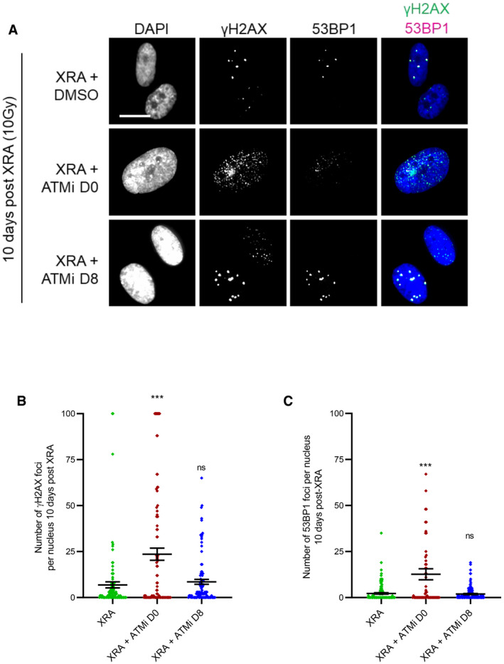
Figure EV3. Impact of ATM inhibitor on persistent DNA damage foci.

-
A–CHCA2‐hT or BJ cells were irradiated with 10 Gy of XRA. Ku‐55933 (ATMi, 5 μM) was added 1 h before XRA (ATMi D0) or 8 days post‐XRA (ATMi D8). (A) BJ cells were fixed 10 days post‐XRA and analyzed for γH2AX or 53BP1 immunofluorescence. Images of γH2AX (green) or 53BP1 (red) and DAPI staining. (B) Quantification of the number of γH2AX foci per nucleus (n = 150). (C) Quantification of the number of 53BP1 foci per nucleus (n = 150). Means ± SEM of foci per nucleus are representative of three independent experiments. Unpaired t‐test: ***P < 0.001, ns: non‐significant.
