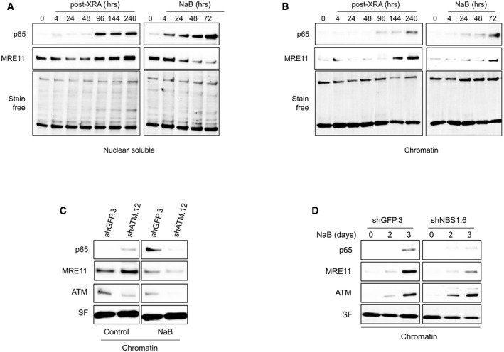
Figure EV5. Chromatin localization of p65 and MRE11 in senescent BJ cells.

-
A, BCells were untreated (time 0), irradiated with 10 Gy of XRA, or treated with 5 mM of NaB. At the indicated time, BJ cells were collected and proteins from the (A) nuclear soluble or (B) chromatin fractions were isolated by subcellular fractionation. Expression of MRE11 or p65 was assessed by Western blot. Stain‐free (SF) imaging of the membrane was used as loading control.
-
CBJ cells infected with lentiviruses expressing shGFP.3 or shATM.12 were treated with 5 mM of NaB for 3 days. Cells were collected and protein from chromatin fraction was isolated by subcellular fractionation. Expression of p65, MRE11, and ATM proteins in chromatin fractions was analyzed by Western blot. Stain‐free (SF) imaging of the membrane was used as loading control.
-
DBJ cells infected with lentiviruses expressing shGFP.3 or shNBS1.6 were treated with 5 mM of NaB. At the indicated times, cells were collected protein from chromatin fraction were isolated by subcellular fractionation. Expression of p65, MRE11, and ATM proteins in chromatin fractions was analyzed by Western blot. Stain‐free (SF) imaging of the membrane was used as loading control.
Source data are available online for this figure.
