-
A, B
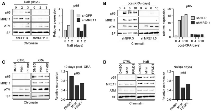
BJ cells infected with lentiviruses expressing shGFP.3 or shMRE11.5 were treated with (A) NaB (5 mM) or (B) 10 Gy of XRA. At the indicated times, cells were collected and protein from chromatin fractions was isolated by subcellular fractionation.
-
C, D
HCA2‐hT cells were (C) irradiated with 10 Gy and at 8 days post‐XRA, Mirin (5 μM).or PMF01 (5 μM) were added for 2 days or (D) cells were treated with NaB (5 mM) +/− Mirin or PFM01 added 24 h after NaB. Alternatively, non‐irradiated HCA2‐hT cells were treated with DMSO, Mirin or PFM01 for 2 days. Ten days post‐XRA or after 3 days of NaB treatment, cells were collected and protein from the chromatin fraction were isolated.
Data information: (A–D) Expression of p65, MRE‐11 and ATM were analyzed by Western blot. Stain‐free (SF) imaging of the membrane was used as loading control. For each panel, expression of p65 were quantified using Image Lab™ (Bio‐Rad) and data were normalized using the total protein quantification from the SF imaging. Protein expression at time 0 or in control was used as baseline. These data are representative of at three independent experiments.

