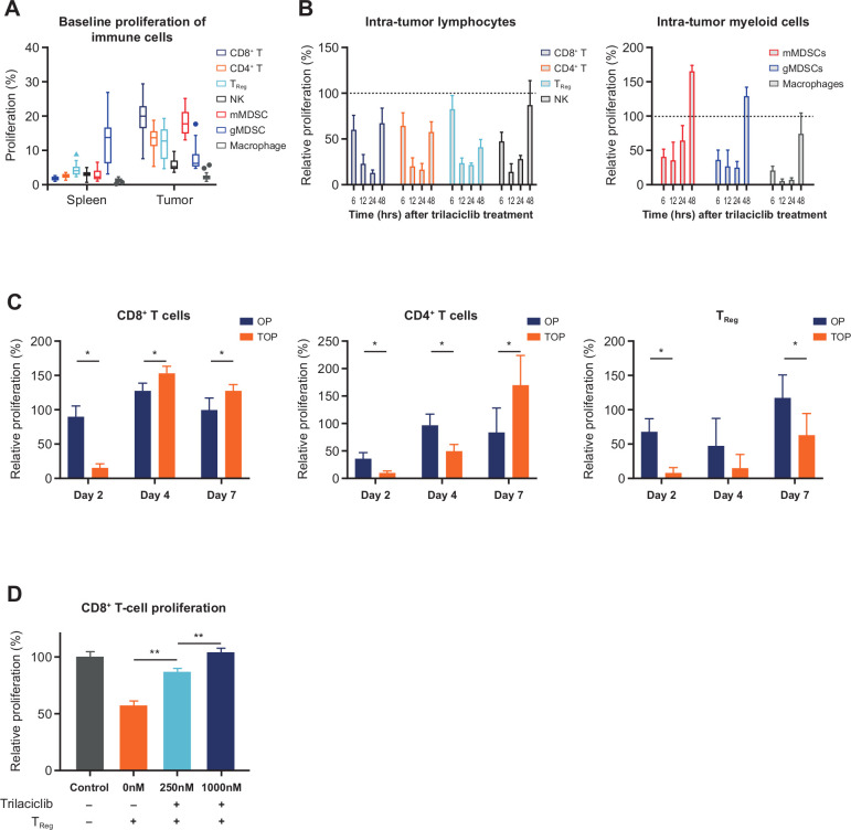
Figure 2.
Addition of trilaciclib to OP treatment combination resulted in transient proliferation arrest followed by a faster recovery of CD8+ and CD4+ T cells compared with Tregs. (A) Baseline percent proliferation status of immune cell populations in spleen and tumors in MC38 tumor-bearing mice (n=16 biological replicates), and proliferation of intratumor lymphoid and myeloid immune cell types at (B) 6–24 hours (n=4 biological replicates) and (C) days 2, 4 and 7 (n=4 or 5 biological replicates) after trilaciclib treatment. Percent proliferation was defined as proportion of EdU+ cells, and relative proliferation was defined as (% EdU+ cells in trilaciclib-treated samples)/(% EdU+ cells in vehicle-treated samples) x 100. Each biological replicate consists of a pool of 3 animals. (D) Proliferation of CD8+ T cells in coculture with trilaciclib-treated Tregs (n=3 independent experiments, each performed with three biological replicates per culture condition). Data represent mean±SD. *p<0.05; **p<0.01. EdU, 5-ethynyl-2ʼ-deoxyuridine; gMDSC, granulocytic myeloid-derived suppressor cell; mMDSC, monocytic myeloid-derived suppressor cell; NK, natural killer; OP, oxaliplatin plus anti-programmed death-ligand-1; TOP, trilaciclib plus oxaliplatin and anti-programmed death-ligand-1; Treg, T-regulatory cell.

