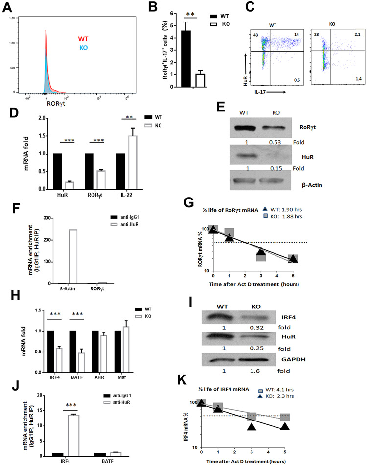
Figure 3. Knockout of HuR reduces RORγt and IRF4 expression in MOG activated Th17 cells.
(A to C) Flow cytometric analysis of RORγt in MOG-activated WT and HuR KO Th17 cells before transferring to Rag1−/− mice. (D, E) The expression of Rorγt mRNA and RORγt protein in WT and HuR KO CD4+ T cells cultured under Th17-cell polarizing condition were examined by RT-qPCR (D) and Western blot (E) analyses, respectively. (F) RNP immunoprecipitation (RIP) followed by RT-qPCR analysis was performed to examine the relative amount of mRNA associated with HuR protein. B-actin mRNA that associated with HuR protein served as the positive control of RIP assay. (G) The relative amount of Rorγt mRNA in WT and HuR KO Th17 cells treated with actomycin D were evaluated by RT-qPCR analysis. (H, I) The relative of Irf4 mRNA (H) and IRF4 protein (I) in MOG-activated WT and HuR KO CD4+ T cells were examined by RT-qPCR and Western blots, respectively. (J) RIP assay showed that Irf4 mRNA was associated with HuR protein in WT Th17 cells. (K) The relative amount of Irf4 mRNA in WT and HuR KO Th17 cells treated with actomycin D were evaluated by RT-qPCR analysis. ** p<0.01, *** p<0.001.

