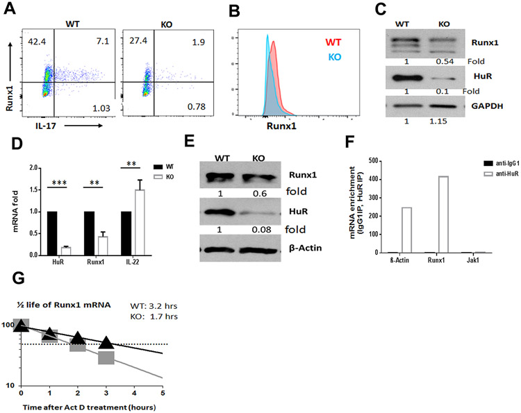
Figure 4. HuR promotes RUNX1 expression in Th17 cells.
(A to D). The expression of RUNX1 in MOG-activated WT and HuR KO CD4+ T cells was evaluated by flow cytometry (A, B) and Western blot analysis (C). (D, E) Levels of Runx1 mRNA (D) and RUNX1 protein (E) in HuR KO CD4+ T cells compared with WT CD4+ T cells under Th17 cell-polarizing condition. (F) RIP assay was used to measure the enrichment in Runx1 mRNA in HuR IP complexes relative to IgG complexes. (G) Actinomycin D treatments were used to assess the half-life of Runx1 mRNA in WT and HuR KO Th17 cells. ** p<0.01, *** p<0.001.

