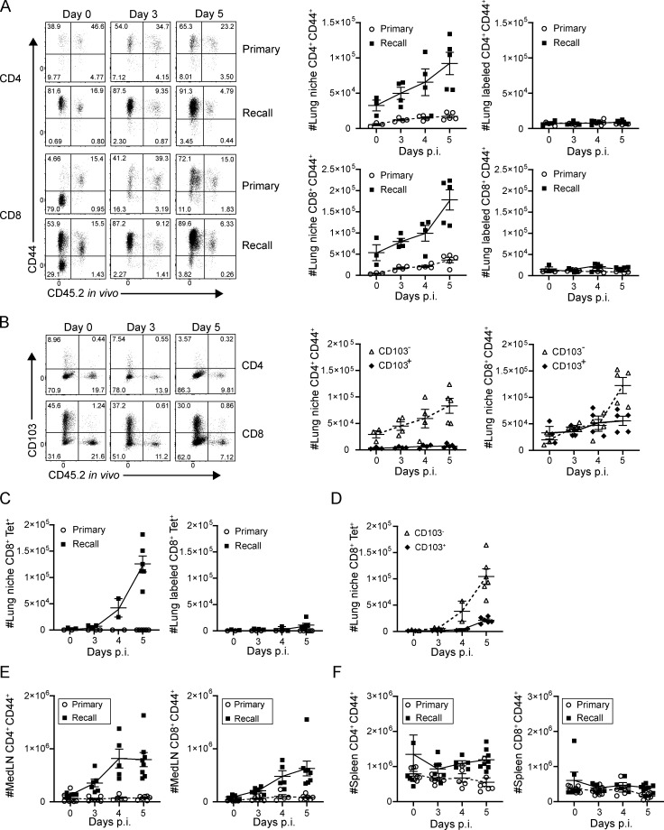
Figure 2.
Tissue-localized protection to heterosubtypic challenge is mediated by rapid lung niche T cell expansion. Lung T cells were isolated from naive and memory mice following in vivo antibody labeling at the indicated days post-infection (p.i.), as in Fig. 1. (A) Accumulation of CD4+CD44+ (upper) and CD8+CD44+ (lower) T cells in the lung following primary and recall challenge is shown in representative flow cytometry plots (left) and in graphs depicting absolute numbers of T cells in the lung niche (middle) and labeled (right) CD44+ T cells. (B) Accumulation of CD103− and CD103+CD4+CD44+ and CD8+CD44+ T cells in the lung during the recall response is shown in representative flow cytometry plots (left), and graphs show absolute numbers of lung niche CD4+CD44+ (middle) and CD8+CD44+ (right) T cells. Data from A and B are from one experiment with three to five mice per group, representative of three independent experiments. (C) Accumulation of lung niche (left) and lung labeled (right) influenza-specific tetramer+ (NP-Tet+) CD8+ T cells over the course of a primary or recall response. (D) Accumulation of CD103− and CD103+CD8+CD44+ NP-Tet+ lung niche T cells during the recall response. Data from C and D were compiled from two independent experiments; n = 2–6 mice/group. (E) Absolute numbers of medLN CD4+CD44+ (left) and CD8+CD44+ (right) T cells during primary and recall responses to PR8 challenge. (F) Absolute numbers of spleen CD4+CD44+ (left) and CD8+CD44+ (right) T cells during primary and recall responses to PR8 challenge. Data from E and F are compiled from three experiments; n = 6–8 mice/group. All error bars show mean ± SEM.

