Figure 2.
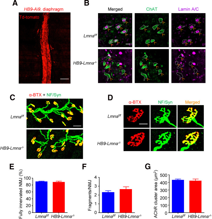
Normal NMJ morphology in adult HB9-Lmna−/− mice. A, Representative images of diaphragm from 2 M HB9::Cre::Ai9 (HB9-Ai9)mouse. Muscles were imaged directly without staining; motor nerves were visibly labeled by td-Tomato signals. Scale bar, 100 µm. B, Representative images of lumbar sections of spinal cords from 2 M control and HB9-Lmna−/− mice. Spinal cord was stained with ChAT (choline acetyltransferase, visualized by Alexa Fluor 488, green) and lamin A/C antibodies (visualized by Alexa Fluor 647, purple). Scale bar, 20 µm. Lamin A/C was deleted from ChAT-positive motoneurons in HB9-Lmna−/−mice (yellow arrows). C, Representative images of 2 M control and HB9-Lmna−/− TA muscles. Muscles were stained with CF568 conjugated α-BTX (red) and anti-NF/Syn antibodies (visualized by Alexa Fluor 488, green). Scale bar, 50 µm. D, Enlarged images of individual NMJ form muscles of the indicated genotype. Scale bar, 20 µm. E–G, Quantification of data in C, D showed unaffected NMJ innervation (E), fragmentation (F), and AChR area (G) in control and mutant mice. Data were shown as mean ± SEM, t(8) = 0.4633, p = 0.6555 for C; t(8) = 1.061, p = 0.3195 for D; t(8) = 0.3450, p = 0.7390 for E; unpaired t test, n = 5 mice per genotype.

