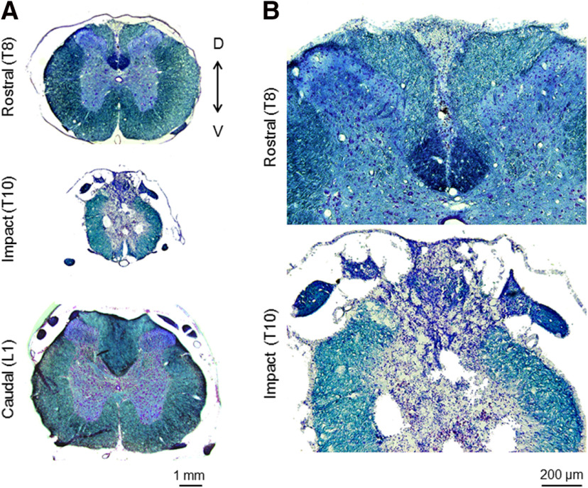
Figure 1.
Spinal cord contusion. Representative sections of the spinal cord taken from a SCI rat at three months postinjury and stained with luxol fast blue and cresyl violet. A, Sections of the spinal cord taken at a rostral site (T8), at the impact site (T10), and at a caudal site (L1). Magnification, 4×. B, Sections of the rostral and impact sites shown at 10× magnification.

