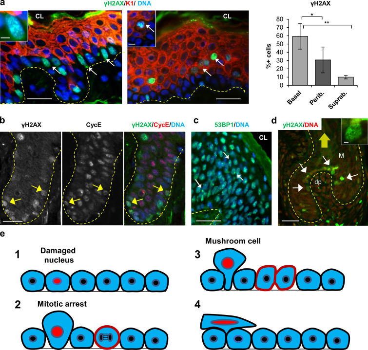
Figure 10.
DNA damage signaling is detected in the squamous epithelia of normal skin. (a) Double IF for γH2AX (green) and K1 (red). Note that γH2AX is usually detected in patches of basal proliferative cells (left panel, arrows) or stratifying differentiating cells (right panel; arrows). γH2AX is also detected in mushroom peribasal cells detaching from the basal layer, expressing K1 (inset in right panel). Amplified inset in left panel shows γH2AX foci. Bar histogram shows the percentage of γH2AX-positive cells in basal, peribasal (Perib.), or suprabasal (Suprab.) layers. (b) Double IF for γH2AX (green) and cyclin E (CycE; red), separate channels and merge as indicated. Arrows point at basal cells strong for γH2AX. (c) IF for 53BP1 (green). 53BP1 bodies are evident in peribasal and suprabasal layers. (d) Detection of γH2AX in the hair follicle germinal matrix (arrows) where keratinocytes are hyperproliferative (DAPI in red). White arrows indicate strong γH2AX cells; yellow arrow indicates the direction of the hair shaft where differentiating cells are incorporated to the hair. Broken lines indicate basement membrane. CL, cornified layer; Dp, dermal papilla; M, matrix. Scale bar, 20 µm; inset scale bar, 3 µm. Nuclear DNA by DAPI (blue). In d, DAPI was made red for better visualizing; γH2AX in green. Quantifications in a are mean ± SEM of 100–160 cells from three fields per each section of three different individuals. Datasets were compared by an unpaired t test (two-sided). *, P < 0.05; **, P < 0.01. Scale bar, 50 µm; inset scale bar, 6 µm, left panel, and 5 µm, right panel. (e) Model for automatic cleansing of squamous epithelia. Basal cells in the epidermis are tightly packed and diving cells have no space. DNA-damaged cells due to active proliferation and RS as a DDR (1), block in mitosis by checkpoints and become larger due to prolonged mitotic arrest and (2) due to lateral forces by more adherent neighbor cells when a new cell division occurs even at a distant spot, are pushed (mushroom cell; Régnier et al., 1986) and spilled into suprabasal layers (stratification; 3). For every stratifying cell (4), one cell will be shed from the surface of the epidermis, thus maintaining self-renewal homeostasis (see also Video 1).

