Figure 7.
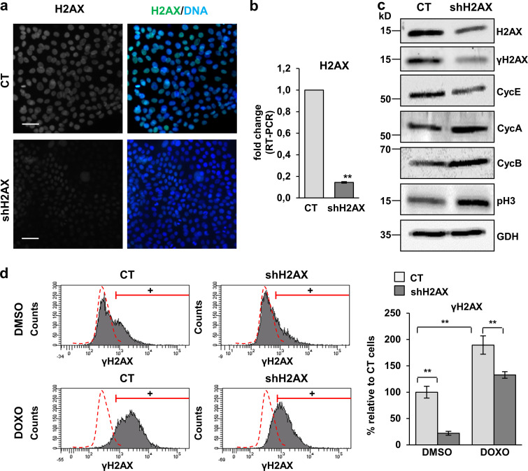
Silencing of H2AX in human keratinocytes induces proliferative markers. (a and b) Loss of expression of total H2AX in primary keratinocytes infected with an shRNA against H2AX (shH2AX) or with the corresponding empty control vector pLKO1 (CT), by IF (a) or by real-time (RT) PCR (b, fold change with respect to control empty vector, CT). Data are mean ± SEM of triplicate samples. **, P < 0.01. (c) Detection of total H2AX, γH2AX, cyclin E (CycE), cyclin A (CycA), cyclin B (CycB), and p-histone H3 (p-H3) by WB in cellular protein fractions of shH2AX or CT cells, GAPDH (GDH) as loading control. (d) Expression of γH2AX in CT or shH2AX keratinocytes treated for 48 h with DMSO (top) or DOXO (bottom), as analyzed by FC (+, positive keratinocytes according to negative isotype antibody control: red broken line). Bar histogram displays the percentage of keratinocytes positive for γH2AX relative to CT. Data are mean ± SEM of duplicate (d) or triplicate (b) samples. Datasets were analyzed by an unpaired t test (two-sided). **, P < 0.01. Scale bar, 50 µm. Nuclear DNA by DAPI. RT-PCR, real-time PCR.

