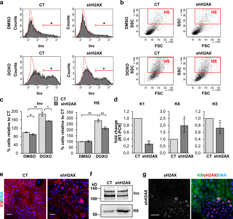
Figure 9.
Inhibition of the γH2AX signaling in human keratinocytes inhibits squamous differentiation and the squamous phenotype. (a) Expression of Inv in CT or shH2AX cells treated for 48 h with DMSO or DOXO, by FC (+, positive keratinocytes according to negative isotype antibody control: red broken line). (b) Light scatter parameters of CT or shH2AX keratinocytes treated for 48 h with DMSO or DOXO, by FC. Red box represents cells displaying HS. (c) Percentage of keratinocytes expressing Inv (left) or displaying HS (right) relative to CT keratinocytes. (d) Expression of K1, K8, and K5 in CT or shH2AX keratinocytes, by real-time (RT) PCR (fold change with respect to CT). (e) Expression of K5 (red) in CT or shH2AX keratinocytes, by IF. (f) Expression of Inv and K8 in CT or shH2AX keratinocytes by WB in insoluble protein fractions of cells in a (same number of cells loaded per lane), by IF. (g) Expression of K8 (green) and γH2AX (red) in unselected shH2AX keratinocytes after infections with CT or shH2AX vectors, by IF. Note that expression of K8 in the colonies is excluding with γH2AX. Nuclear DNA in blue by DAPI. Data are mean ± SEM of duplicate (c) or triplicate (d) samples. Datasets were compared by an unpaired t test (two-sided). *, P < 0.05; **, P < 0.01. Scale bar, 50 µm. FSC, forward scatter; SSC, side scatter.

