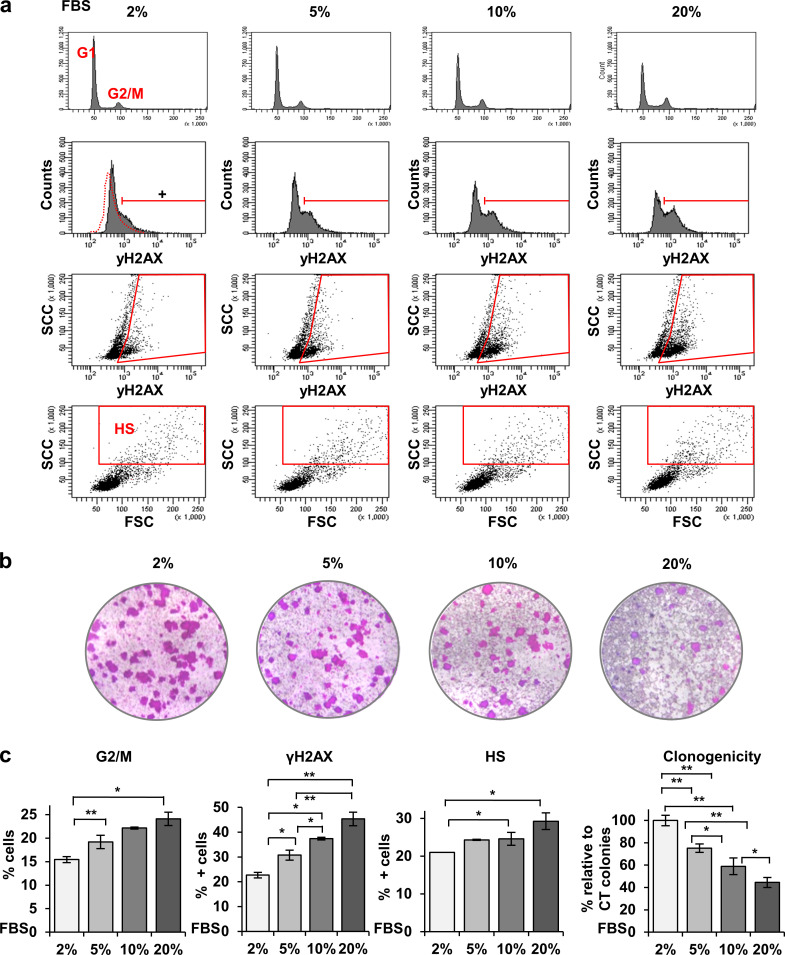
Figure S3.
Increasing serum concentration induces γH2AX in human keratinocytes and results in loss of proliferative potential. (a) FC analyses of primary keratinocytes cultured in increasing serum concentrations as indicated, from top to bottom for DNA content by PI, γH2AX (DNA damage), γH2AX versus light side scatter (SCC), SCC versus forward scatter (FSC). Red gate is for γH2AX-positive cells according to negative isotype control, or cells displaying HS typical of squamous differentiation. (b) Clonogenicity assays of keratinocytes cultured in increasing serum concentrations for 9 d. Figure shows representative images of triplicate samples. (c) Quantifications of cells in the G2/M phase of the cell cycle, positive for γH2AX, or displaying HS, according to gates in a. Right last histogram displays number of growing colonies relative to 2% serum in the culture medium. Data are mean ± SEM of triplicate samples. Datasets were compared by an unpaired t test (two-sided). *, P < 0.05; **, P < 0.01. This figure complements Fig. 1.

