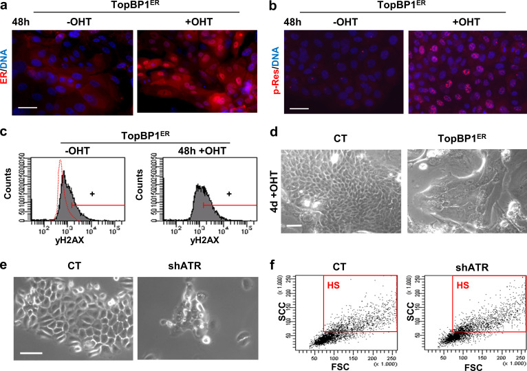
Figure S4.
Gain or loss of ATR activity affects terminal keratinocyte differentiation. (a and b) Expression of estrogen receptor (ER, red, a) or p-Res (red, b) in keratinocytes infected with control vector pMXPIE (CT) or with the TopB1PER construct, 48 h in the absence or presence of OHT. (c) γH2AX expression in keratinocytes as in a, by FC (+, positive keratinocytes according to negative isotype antibody control). (d) Phase contrast images of cells in a after 4 d in the presence of OHT, as indicated. (e) Phase contrast images of keratinocytes 4 d after infection with specific shATR or with the corresponding control vector plKO.1 (CT). (f) Light scatter parameters of keratinocytes 4 d after infection with shATR or with CT. Red box gates cells with HS typical of terminal differentiation. Scale bar, 50 µm. This figure complements Fig. 3 and Fig. 4. FSC, forward scatter; SSC, side scatter.

