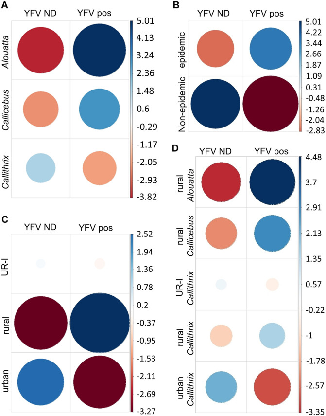
Fig 2. Association between detection of yellow fever virus (YFV) in the liver of non-human primate (NHP) carcasses with (a) NHP genera, (b) period of sampling (c) environment, and (d) environment plus NHP genera.
YFV ND: yellow fever virus RNA was not detected. YFV pos: yellow fever virus RNA was detected. Pearson residuals (standardized) were extracted from the chi-square function and plotted. In each cell, the size of the circle is proportional to the amount of cell contribution, and colors indicate positive residuals (blue) or negative residuals (red). The vertical bars indicate Pearson residuals values. Analyses were run in R software v.3.6.0.

