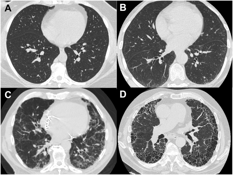
Figure 2. Representative Images from Cohort Subjects.
A. High-resolution CT (HRCT) image of the chest from a study subject whose scan was read as normal, without signs of interstitial lung disease or fibrosis. B. HRCT image from subject who was categorized as having “Probable Fibrotic ILD.” C. Representative HRCT image from subject who was characterized as having “Definite Fibrotic ILD.” D. HRCT image from a case of previously diagnosed, established Idiopathic Pulmonary Fibrosis (IPF) in one of the study families.

