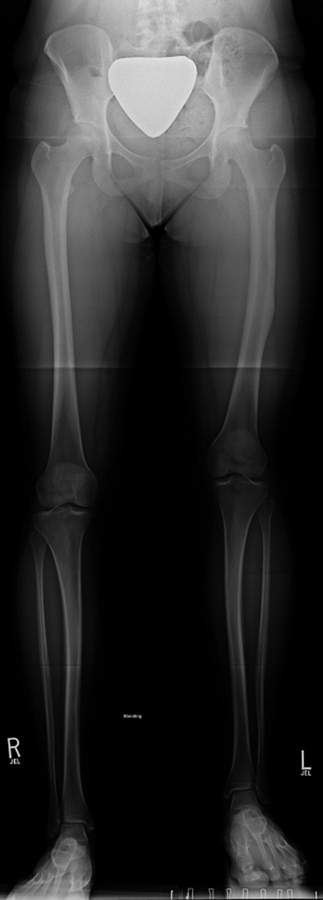
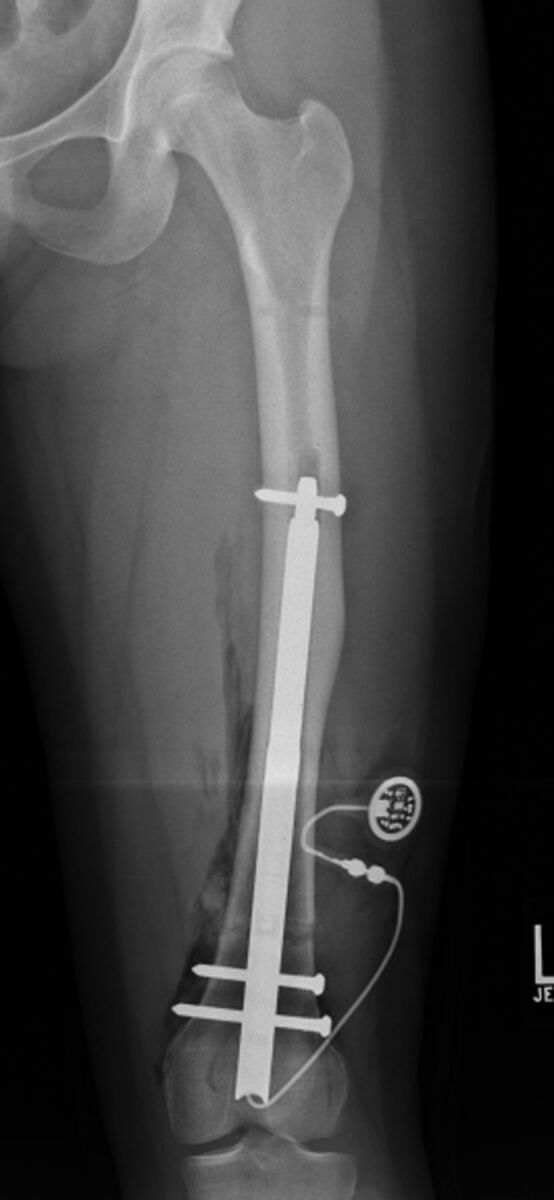
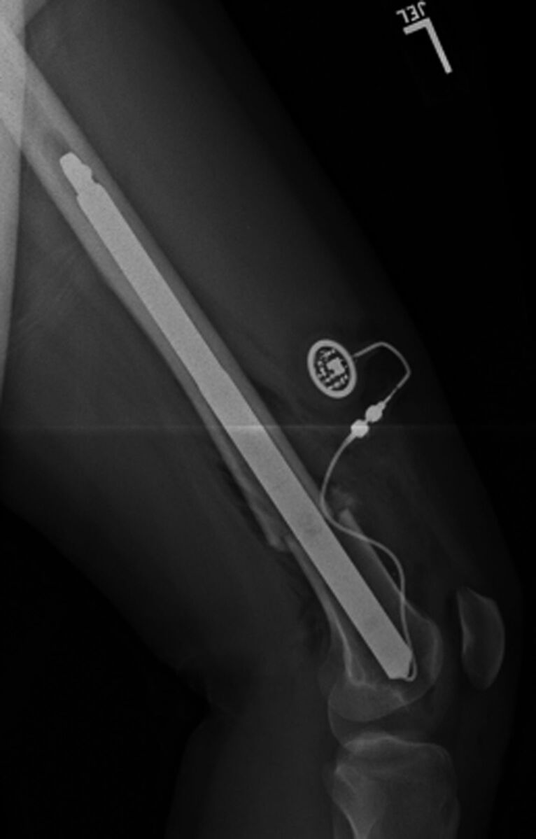
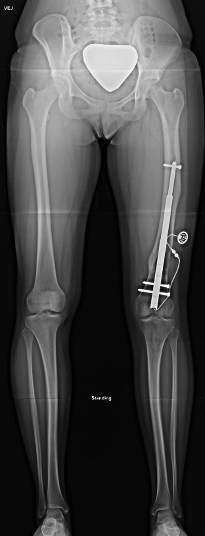
Figs. 1-A through 1-D Illustrative case of a patient treated with a FITBONE TAA motorized intramedullary nail inserted in a retrograde femoral fashion. The patient experienced no complications during treatment.
Fig. 1-A.

Preoperative anteroposterior radiograph of the lower extremities made with the patient standing on a 4.5-cm block under the left foot. Note the midshaft femoral varus deformity and the midshaft tibial valgus deformity.
Fig. 1-B.

Anteroposterior radiograph of the femur made at the end of surgery. Note the retrograde insertion, proximal and distal locking bolts, and the cable leading from the base of the nail through a drill-hole in the lateral femoral condyle. This cable is connected at the time of surgery to the subcutaneously implanted radiofrequency receiver. To effect lengthening, radiofrequency waves are transmitted transcutaneously to the receiver, which converts them to electrical impulses discharged to the electric motor housed within the nail.
Fig. 1-C.

Lateral radiograph of the femur made at the end of surgery. Note the straight nail. During preoperative planning and intraoperative canal preparation by reaming, the diameter, length, and straight contour of the nail must be taken into consideration.
Fig. 1-D.

Anteroposterior radiograph of the patient made six months after surgery. The limb lengths were equal. The patient had excellent clinical and radiographic alignment of the legs and had returned to full function. The nail was removed, per manufacturer’s recommendation, eighteen months after installation.
