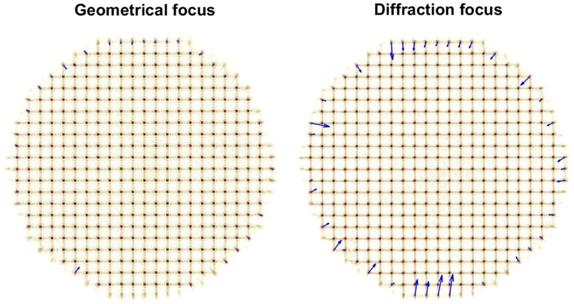
Fig. 3.

Sequence of SHWS images captured with the experimental setup in Fig. 2 while the iris is closing and with the pixelated detector at the geometrical focus (left) and diffraction focus (right) of the lenslet array (see Visualization 1). The blue arrows show the spot displacements from their nominal position, magnified 20 times for display purposes. The displacements of outermost spots, which correspond to partially illuminated lenslets, are substantially larger at the diffraction focus.
