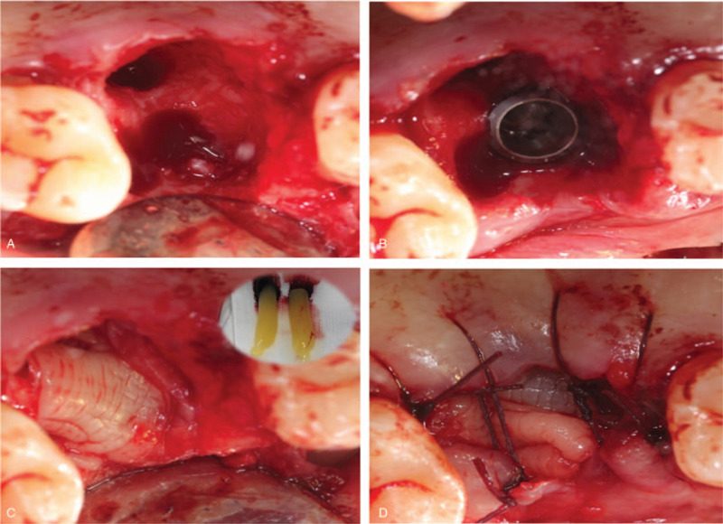
Figure 2.

Intraoperative photographs and illustration describing each step of the surgery. (A) The tooth was carefully removed; (B) A bone defect gap around the implant; (C) 2 PRF membranes covered over the bone materials; (D) Semi-open flap was used. PRF = platelet-rich fibrin.
