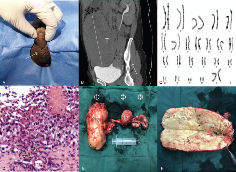
Figure 1.

Clinicopathological features. A. Physical examination reveals a urethral fistula on the ventral surface (arrow). B. Computerized tomography shows a large tumor (T, B). C. Karyotype analysis of the patient. D. Photomicrograph of biopsied tissue shows seminoma. E. Image shows the tumor (①), uterus (②), and tubo-ovarian nodule (③). F. Image shows specimen bisection and extensive necrosis.
