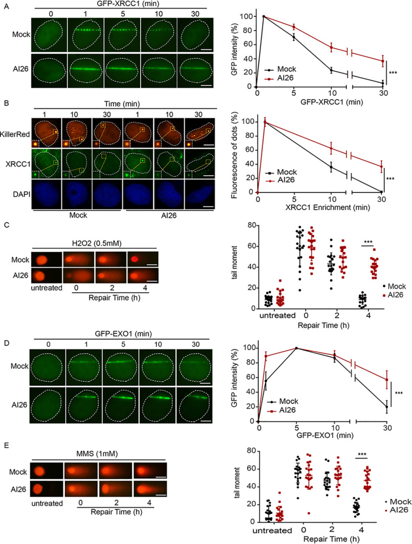
Figure 4.
AI26 treatment impairs DNA damage repair. A and B, AI26 treatment traps XRCC1 at DNA lesions. U2OS cells expressing GFP-XRCC1 were pretreated with or without 10 μm AI26 for 1 h, and the retention of XRCC1 at laser strip was examined with live cell imaging (A). U2OS cells expressing KillerRed were pretreated with or without 10 μm AI26 for 1 h, and the endogenous XRCC1 at the oxidative damage sites was examined by IF with anti-XRCC1 antibodies. The scale bar represents 5 μm. B, results are displayed as means ± S.D. from 50 cells (n = 3 independent experiments). ***, P < 0.001. The scale bar represents 5 μm. C, AI26 treatment suppresses SSBR. U2OS cells were pretreated with or without 10 μm AI26 for 1 h, followed by 0.5 mm H2O2 for 5 min. Alkaline comet assays were performed to examine the rate of SSBR in time course experiments. The tail moments were determined from at least 50 cells at each time point in each experiment, and three independent experiments were carried out. ***, P < 0.001. The scale bar represents 30 μm. D, AI26 treatment traps EXO1 at DNA lesions. U2OS cells expressing GFP-EXO1 were pretreated with or without 10 μm AI26 for 1 h, and the retention of EXO1 at laser strip was examined with live cell imaging. Three independent experiments were carried out. ***, P < 0.001. The scale bar represents 5 μm. E, AI26 treatment suppresses DSBR. U2OS cells were pretreated with or without 10 μm AI26 for 1 h, followed by 1 mm MMS for 30 min. Neutral comet assays were performed to examine the rate of DSBR in time course experiments. The scale bar represents 30 μm.

