
Figure 1.
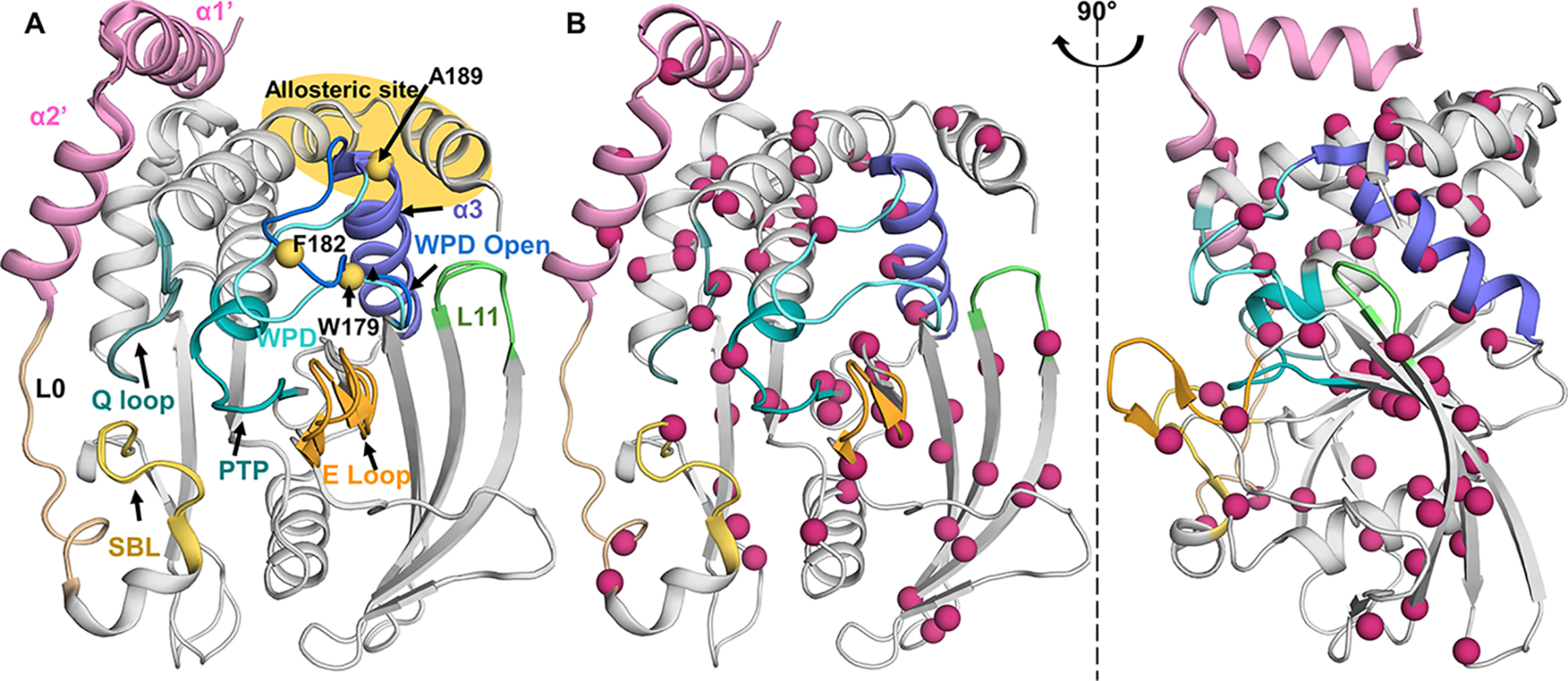
13C ILV methyl groups are well-dispersed throughout PTP1B. A, overlay of PTP1B (open state, PDB 5K9V; closed state, PDB 5K9W); active site: PTP and Q loop (teal), WPD loop (open, blue; closed, cyan); substrate recruitment: E and SBL loop (yellow/orange). Purple, helix α3; green, loop L11. The known allosteric site is highlighted in yellow at the intersection of helices α3, α6, and α7. Residues previously studied by 15N ct-CPMG relaxation measurements are shown as yellow spheres. B, PTP1B (PDB 5K9W) Ile, Leu, and Val residues depicted as pink spheres; well-distributed throughout PTP1B.