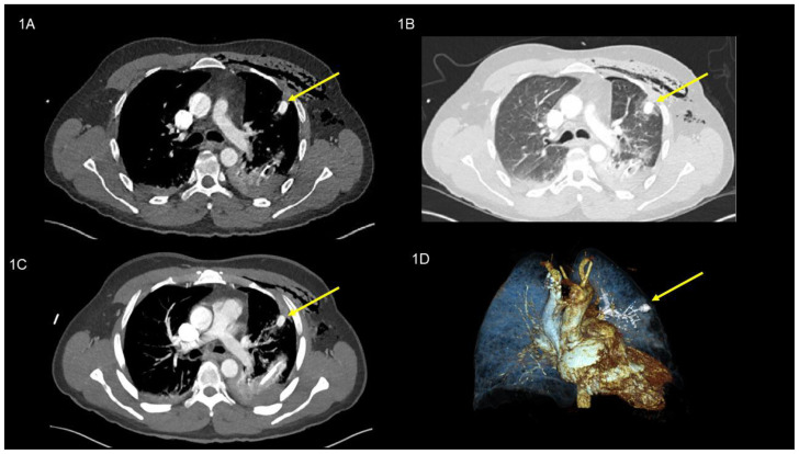
Figure 1.
49-year-old man with peripheral pulmonary artery pseudoaneurysm in the left upper lobe. Initial images after the trauma (all images are at the same level).
Findings:
Axial contrast enhanced chest CT in the dual-phase showed initially hemopneumothorax and extended subcutaneous/intramuscular emphysema and a round lesion (pulmonary artery pseudoaneurysm), yellow arrow, (16 × 11 mm), (135 Hounsfield units; equivalent to contrast agent density), in the left upper lobe (anterior segment S III), (a) axial soft tissue window, (B) axial lung window, (c) MIP 10 mm, and (d) Image reconstruction.
Technique: CT SIEMENS (Somatom Definition Edge 128) 142 mAs, 120kV, 1mm slice thickness, 90 ml Iomeron 400 KM i.v.

