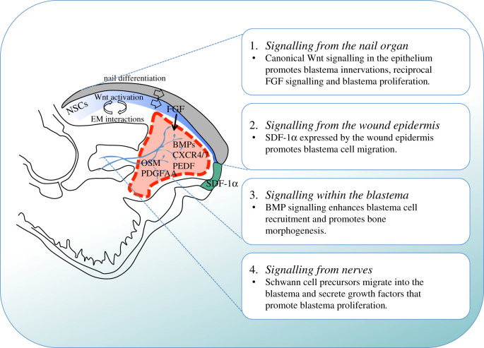
Figure 4.
Known signalling mechanisms that contribute to mammalian digit tip regeneration. Indicated are the signalling mechanisms that originate from the nail organ (1), from the wound epidermis (2), within the blastema (3) and from nerves (4). During distal digit tip amputation the wound site is covered by regenerating nail epithelial cells that arise from the nail stem cells (NSCs). This epithelium activates Wnt signalling that promotes differentiation of NSCs that give rise to the nail plate. Additionally, through epithelial–mesenchymal (EM) interactions, this Wnt signalling promotes blastema innervation, which in turn enables FGF2 expression in the nail epithelium and promotes blastema cell proliferation. Blastema cell migration is stimulated by the cytokine SDF-1α that is expressed by the wound epidermis while the reciprocal receptors CXCR4/7 are detected on blastema cells, together with BMPs and the anti-angiogenic factor Pedf. Schwann cell precursors that arise from injured peripheral nerves migrate into the blastema and secrete growth factors including oncostatin M (OSM) and platelet derived growth factor AA (PDGFAA) that promote blastema proliferation. Nerves are shown in blue; the wound epidermis is indicated in green; the blastema is indicated by the red-hatched line; the nail epithelium is indicated in blue and the nail plate is coloured grey.

