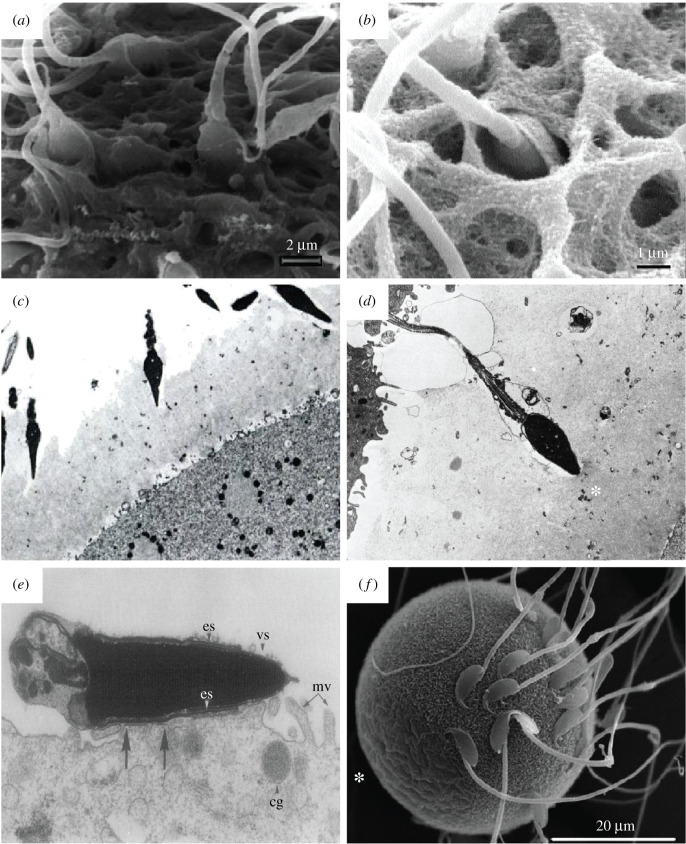
Figure 3.
Conventional EM of mammalian sperm–oocyte interactions. (a) SEM micrograph of a mature human oocyte, showing the vertical binding of the sperm with a penetration of the apical tip of the sperm head into the ZP [33]. (b) SEM micrograph of a human sperm–oocyte interaction, showing the vertical binding of a sperm head vanishing into the the ZP [14]. (c) TEM micrograph of human sperm–oocyte interaction in vitro, showing acrosome-reacted sperm invading the ZP of a polyspermic ovum at different angles (×3330) [34]. (d) TEM micrograph of human sperm–oocyte interaction in vitro, showing an acrosome-reacted sperm that has penetrated about half the thickness of the ZP, just blocked outside the inner surface of the ZP (*) which is denser and more compact than the outer surface, thus depicting the block to polyspermy (×4330) [34]. (e) TEM micrograph showing the fusion of a tubal mouse egg incubated with capacitated epididymal sperm for 60 min. It shows the fusion of the sperm at the postacrosomal cap of the equatorial segment (es); cg, cortical granules; mv, microvilli; vs, vesiculated plasma and outer acrosomal membranes (×31 200) [35]. (f) SEM micrograph of a wild-type mouse egg incubated with sperm for 25 min, clearly showing sperm are not bound to microvilli-free region (*) [36].

