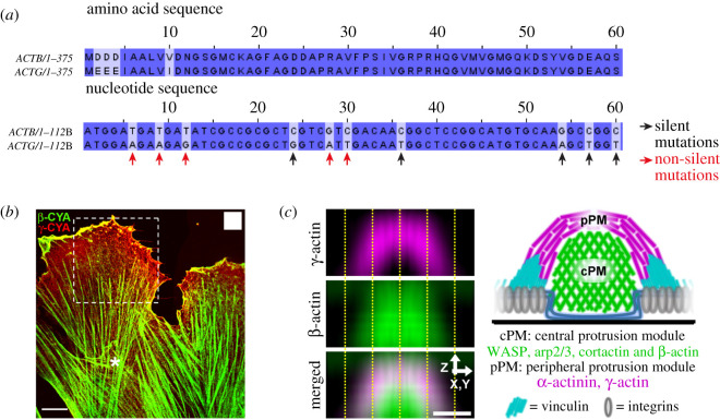
Figure 4.
Evidence that highly similar cytoplasmic actins can nevertheless assemble into functionally different actin networks in cells. (a) Beginning of the nucleotide and amino acid sequence of β- and γ-actins. These proteins only differ in four amino acids located at the N-terminal end, although their nucleotide sequences have a much higher number of silent mutations (e.g. black arrows). (b) Example of the differential localization of β- (green) and γ-(red) actins at the cell scale, in migrating human subcutaneous fibroblasts (adapted from [54], scale bar, 10 µm) (c) Another example of the differential localization of β- (green) and γ-actins (magenta) within a same structure, podosomes (adapted from [57], scale bar: 0.5 µm). A linear network of γ-actin is surrounding a branched β-actin core.

