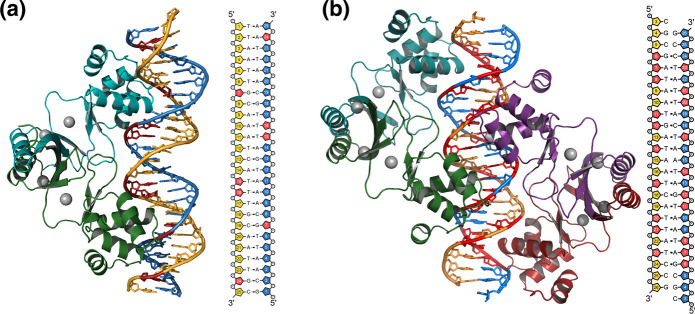
Fig. 7.
Homology models of AsFur in the two Fur-DNA interaction modes observed for MgFur and reported by Deng et al. (2015). a A dimer of AsFur interacting with the feoAB1 operator. b Two AsFur dimers interacting with the E. coli Fur box. Each AsFur monomer is coloured individually (monomer A, dark green; monomer B, turquoise; monomer C, purple; monomer D, red) and the DNA strands are coloured in dark yellow and blue for the primary and complementary strands, respectively. Nucleotides coloured in red indicate base contacts with AsFur. The modelled Mn2+ ions are indicated as grey spheres. (Color figure online)

