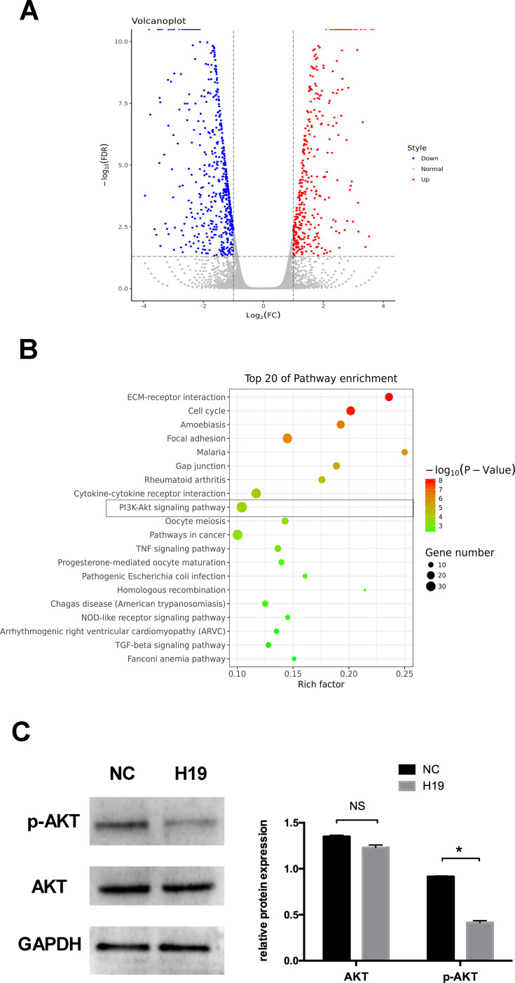
Figure 4.
RNA sequencing following transfection with H19 vector for 48 h. (A) Volcano plot of differentially expressed mRNAs in the control and H19 groups. Red points: upregulated mRNAs; blue points: downregulated mRNAs. (B) The top 20 enrichments in the KEGG pathway analysis of differentially expressed genes. (C) Western blot analyses of p-AKT and AKT after PDLCs transfected with H19 vector for 48 h. Histograms show quantification of the band intensities (Analysis of variance *P <0.05; NS: non-significance, P >0.05; n=9 replicates derived from three donors).

