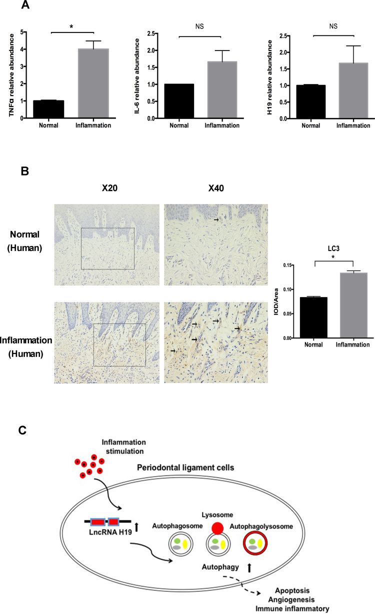
Figure 6.
The levels of autophagy and H19 were increased in periodontitis-affected human gingival tissue. (A) Relative RNA expression of H19, TNFα, and IL-6 in the human gingival tissue with or without periodontitis. (B) Immunohistochemical staining of LC3 in healthy and inflamed human gingival tissue. Black arrows identified the LC3 staining of gingival tissue of periodontitis patients. Quantitative analysis showed the LC3 staining in inflammation group was significantly stronger than the control group. (C) Schematic diagram showing the regulation of autophagy by H19 in PDLCs (Analysis of variance *P <0.05; NS: non-significance, P >0.05; n=10 replicates derived from 5 donors in each group).

