Figure 6.
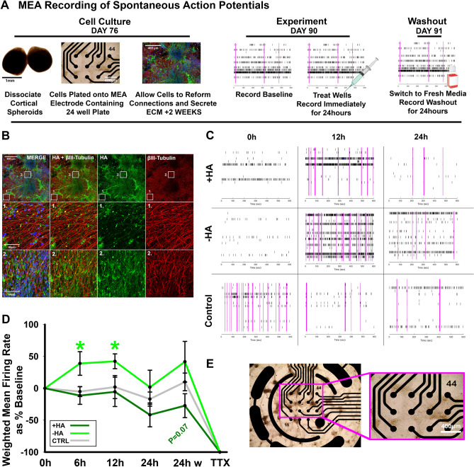
HA regulates spontaneous action potential formation. (A) Workflow for MEA experiments. Day 76 spheroids are dissociated enzymatically and plated onto 24-well microelectrode array plates. After 2 weeks, neurons have re-established neural networks and the HA-based ECM. At day 90, the cells are treated and spontaneous action potentials are recorded for 10 min/hour for 24 h. After 24 h, fresh media is added to wells for a washout recording of 10 min/hour for 24 h. (B) Representative confocal image for βIII-tubulin (neurons, red), DAPI (nuclei, blue), and HABP (HA, green) for cortical spheroids dissociated at day 76 and imaged at day 90. Middle and bottom panels highlight detail of HABP and βIII-tubulin area. Scale bar top panel: 400 μm, middle and bottom panel: 60 μm. (C) Raster plots of spontaneous action potentials in + HA (top), − HA (middle), and control wells (bottom) at 0 h, 12 h, and 24 h after treatment. Each black bar is a single spike, meaning one electrode has fired, pink bars highlight network bursts, where more than one electrode detect simultaneous activity. High network bursting is characteristic of hyperexcitable networks. (D) Quantification of the weighted mean firing rate (WMFR) for each treatment at 0 h, 6 h, 12 h, 24 h post treatment, 24 h post washout, and immediately after TTX treatment. Hyaluronidase treatment significantly increased WMFR at 6 h and 12 h following treatment. (E) Brightfield image of dissociated spheroids plated for 2 weeks on a microelectrode array. Inset shows association of neurons with electrodes. Scale bar 400 µm.

