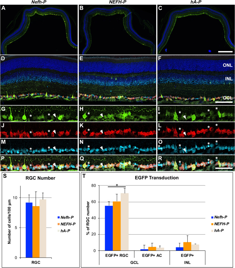
Figure 4.
EGFP expression following IV delivery of Nefh-P, NEFH-P or hA-P. Nefh-P, NEFH-P or hA-P was delivered via IV injection (1 × 10⁹) to adult wild type mice (n = 4). Four weeks post-delivery, eyes were enucleated, fixed in 4% pfa and cryosectioned. Immunocytochemistry for EGFP (Alexa-Fluor-488 label, green), RBPMS (Cy3 label, red) and PAX6 (Cy5 label, light blue) was carried out. (A–C) Overview of EGFP-transduced retinas. (D–F) Overview of EGFP-, RBPMS- and PAX6-label in EGFP-transduced retinas. Distribution of EGFP (G–I), RBPMS (J–L), PAX6 (M–O) and their overlay (P–R) in the GCL is presented. Most cells were positive for the three markers demonstrating that they were EGFP-transduced RGCs (G–R). Arrowheads indicate amacrine cells (ACs, RBPMS negative/PAX6 positive) that expressed EGFP, while asterisk indicate ACs (RBPMS negative/PAX6 positive), which did not express EGFP. Bar chart representation of the number of RGCs in the transduced retinas (S) and the percentage of EGFP-positive RGCs and ACs in the GCL and the EGFP-positive cells in the INL (T); the number of RGCs was taken as 100%; bars represent mean + SD; *p < 0.05 (ANOVA). Automated (for RGC and INL cells) and manual (for ACs) quantification was carried out in cellSens. ONL outer nuclear layer, INL inner nuclear layer, GCL ganglion cell layer. Scale bars: 500 μm (C), 100 μm (F), 50 μm (R).

