
Figure 2.
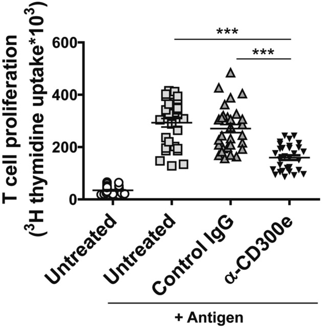
Activation through CD300e compromises the ability of monocytes to activate antigen-specific T cells. Monocytes isolated from 10 patients with melanoma were seeded in triplicate as above on uncoated plates or on plates coated with isotype IgG or anti-CD300e. After 24 h, cells were harvested and co-cultured with CD4+ T lymphocytes purified from the same patients in presence of specific melanoma antigen (MAGE-A3). Lymphocyte proliferation was measured by 3[H]-TdR incorporation. Data are shown as mean ± SEM of 10 independent experiments. Significance was determined by Student’s t-test. ***p ≤ 0.001.