Figure 7.

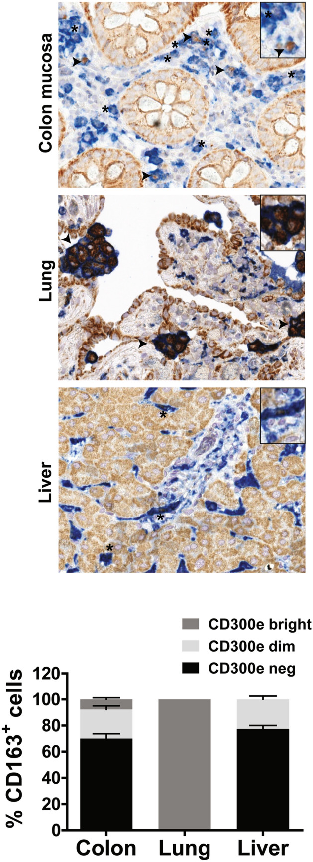
Expression of CD300e in tissue macrophages. Immunohistochemical staining for CD300e (brown) and CD163 (blue) was performed on sections of normal colon mucosa, lung and liver. Arrows highlight macrophages which strongly express CD300e, while asterisks highlight weakly expressing macrophages. The percentage of CD163+ macrophages, CD300ebright, CD300edim or not expressing CD300e, in 3 patients for colon mucosa and liver and in 4 patients for lung was calculated and expressed in the bottom plot as mean ± SEM. Original magnification 400× , inset magnification 600 × .
