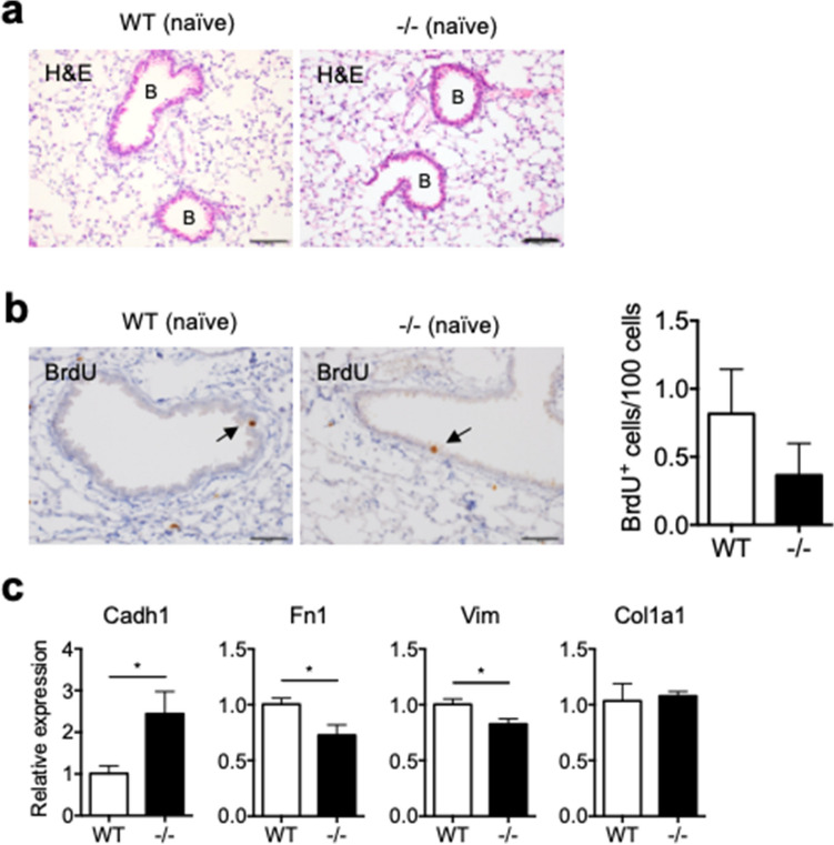
Figure 1.
Characterization of lungs from naïve WT and Spred2−/− mice. (a) Lung tissues were removed from WT and Spred2−/− mice and examined by H&E staining. Bronchi are indicated by (B). The original magnification was 200×. The scale bars are 100 μm. (b) Naïve WT and Spred2−/− mice were intraperitoneally injected with BrdU. Two hours later, mice were euthanized and the incorporation of BrdU was examined by IHC. The number of BrdU+ cells in five bronchi were counted and presented as BrdU+ cells/100 cells. The results are presented as mean ± SEM. n = 5. The original magnification was 400×. The scale bars are 50 μm. (c) The expression of the Cadh1, Fn1, Vim and Col1a1 gene was examined by qRT-PCR. The results are presented as mean ± SD. n = 4. *p < 0.05. ***p < 0.0001.

