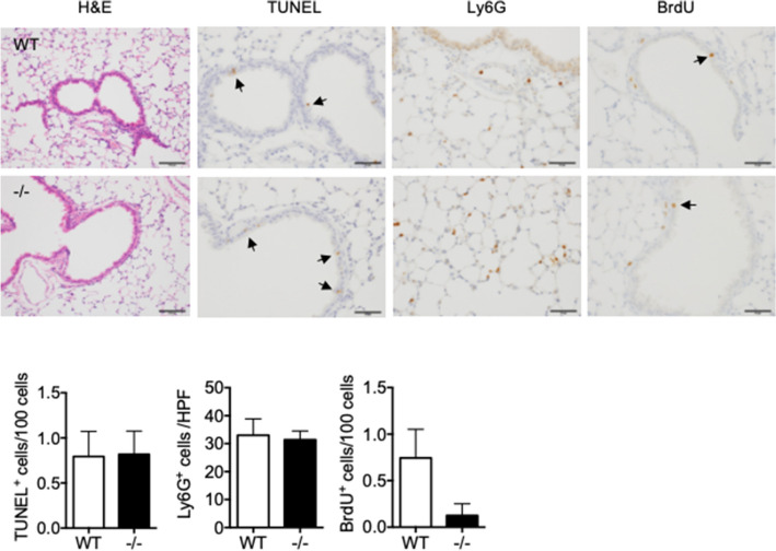
Figure 3.
Characterization of lungs from naïve WT and Spred2−/− mice 1 day after BLM administration. Mice were injected intratracheally with 1.5 mg/kg BLM and lung tissues were harvested after 24 h. Tissue sections were subjected to H&E staining, TUNEL and IHC with anti-Ly6G Ab. To detect the proliferation of cells, BrdU was intraperitoneally injected 2 h before the harvest and the incorporation of BrdU was evaluated by IHC. Representative images are shown. The original magnification was 200 × for H&E staining and 400 × for TUNEL, Ly6G or BrdU staining. Arrows indicate positive cells. The number of TUNEL+ or BrdU+ cells in five bronchi were counted and presented as TUNEL+ or BrdU+ cells/100 cells. Ly6G+ cells in five high power field (400×) were counted and the results are presented as mean ± SEM. n = 5. Scale bars are 100 μm for H&E and Ly6G staining and 50 μm for TUNEL and BrdU staining.

