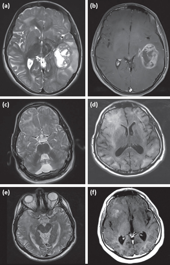
Figure 1.

Brain MRI findings belonging to two patients with L2HGA who had brain tumor; 1 (a, b): brain MRI findings of subject 6; mass compressing the surrounding tissues with heterogeneous contrast uptake and cystic component in the temporoparietal region in the left hemisphere in T2-weighted and contrast-enhanced axial sections 1 (c–e): brain MRI findings of subject 22; 1 (c, d): asymmetrical increased signal intensity in the subcortical and deep white matter in the right frontal region in T2 and FLAIR axial sections dated 2011, 1 (e-f): mass with heterogeneous contrast uptake in the right temporal region in T2 axial section and the frontal regions in the contrast-enhanced section extending to the inferior temporal area dated 2013
