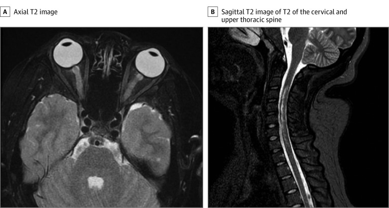
Figure. Neuroimaging Studies at Diagnosis.
A, Axial T2 demonstrating bilateral signal prolongation in the optic nerves. There was no associated postcontrast enhancement of the optic nerves. B, Sagittal T2 of the cervical and upper thoracic spine demonstrating signal patchy signal prolongation throughout the upper spine. On postcontrast imaging, cervical lesions were noted to have patchy and heterogenous signal throughout the cervical cord in the same distribution as T2 signal prolongation.

