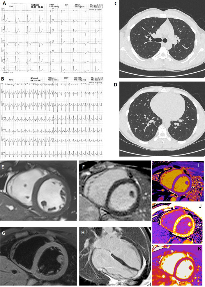
Figure 1.
Instrumental findings in a player with increased troponin I level. In the only SARS-CoV-2-positive player (asymptomatic) with increased troponin I level, resting (A) and stress-test (B) ECG were normal. Chest CT at the level of the plane passing through the upper right lobar bronchus (C) and of the plane passing through lung bases (D) was absolutely normal. (E–M) Cardiac magnetic resonance images acquired using a 1.5 T Siemens Aera (Siemens Healthcare, Erlangen, Germany). (E) Short-axis cine balanced steady-state free precession showed normal left ventricle end-diastolic volume, wall thickness and motion. (F) Short-axis T2 image showed no oedema. (G, H) Short-axis and four-chamber views showed no alteration of late gadolinium enhancement. (I, J) Short-axis T1 native and T1 postcontrast maps showed normal values of T1 and extracellular volume. (K) T2 map showed no oedema.

