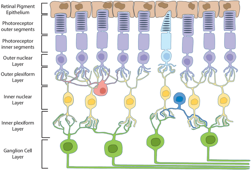
Figure 1. Schematic illustration of retinal cell layers.
RPE cells (brown) maintain close contact with and phagocytose rod (purple) and cone (light blue) photoreceptor outer segments. Bipolar cells (yellow) synapse with and transfer information between photoreceptors and ganglion cells (green). Axons of the ganglion cell layer converge to form the optic nerve. Horizontal (red) and amacrine (dark blue) cells serve multiple functions by integrating and regulating signal transduction throughout the retina.

