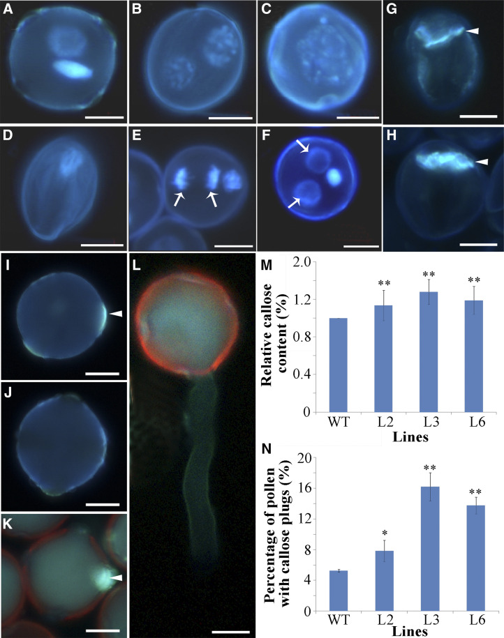
Figure 3.
Overexpression of PSP231 results in aberrant hyper-callose deposition impairing pollen cell division. A to F, Pollen grains of T3-generation transgenic tobacco lines L2, L3, and L6 and the wild type were stained by 4',6-diamidino-2-phenylindole (a nucleus-specific dye that emits stronger fluorescence when bound to DNA). Shown are mature and bicellular pollen of the wild type (control; A), two equal nuclei in transgenic pollen (B), fusion nuclei in transgenic pollen (C), aborted transgenic pollen (D), transgenic pollen with abnormal cell division (E), and two vegetative cell nuclei in transgenic pollen (F). Arrows indicate extra vegetative cell nuclei. G and H, Decolorized aniline blue staining showing the callose wall between the vegetative cell and the germ cell in pollen from wild-type (G) and PSP231 transgenic tobacco (H). I and J, Aniline blue-stained mature pollen from PSP231 transgenic tobacco (I) and the wild type (J). K and L, Aniline blue-stained pollen germinated in vitro from PSP231 transgenic tobacco lines (K) and the wild type (L). M, Statistical analysis of the relative callose content in dividing pollen cells. Relative callose content in the wild type (WT) is set to 1. N, Statistical analysis of the percentage (%) of pollen grains with callose plugs in germ pores in the transgenic lines and wild-type tobacco. For M and N, data are means ± sd for three replicates. Dunnett's t test demonstrated that there was significant difference (*P < 0.05) or very significant difference (**P < 0.01) between the PSP231 transgenic plants and the wild type. Arrowheads in G, H, I, and K indicate callose deposition. Scale bars = 10 μm (A–H) and 2 μm (I–L).

